Knowledge
Magazine
-

The 24-Hour Sleep Model: Why Better Sleep Starts the Morning Before
8 min read
-

Eating for Longevity: Lessons From the World's Oldest People
9 min read
-

The Summer Health Kit: Supplies and Gadgets for Hot Days and Warm Nights
9 min read
Weekly picks on longevity, brands, and health science. No spam—unsubscribe anytime.
Tools
- →The 30s Longevity Guide for Women
- →Postpartum Recovery Guide
- →Perimenopause Longevity Guide
- →Dopamine Reset Guide
- →Pregnancy Longevity Guide
- →Peptide Guide
- →Home Detox Guide
- →Slow Aging Guide
- →Pro Supplement Guide
- →Safe Supplement Guide
- →Bio Age Calculator
- →Blood Testing Guide
- →Deep Sleep Guide
- →Pro Longevity Dashboard
By Category
By Goal
Knowledge
- →What is Longevity?
- →Improve diet
- →More focus
- →Look younger
- →Lose weight
- →Track biomarkers
- →Improve sleep
- →Build muscle
- →Increase cardio
- →Detect diseases
- →Recover faster
- →Relieve stress
Magazine
-
The 24-Hour Sleep Model: Why Better Sleep Starts the Morning Before
8 min read
-
Eating for Longevity: Lessons From the World's Oldest People
9 min read
-
The Summer Health Kit: Supplies and Gadgets for Hot Days and Warm Nights
9 min read
Weekly picks on longevity, brands, and health science. No spam—unsubscribe anytime.
Brands
- →Apps
- →At Home Tests & Kits
- →Events
- →Experts & Clinics
- →Foods & Beverages
- →Gadgets & Equipment
- →Gyms & Workouts
- →Supplements
- →Travel & Retreats
- →Wearables & Trackers
Tools
- →The 30s Longevity Guide for Women
- →Postpartum Recovery Guide
- →Perimenopause Longevity Guide
- →Dopamine Reset Guide
- →Pregnancy Longevity Guide
- →Peptide Guide
- →Home Detox Guide
- →Slow Aging Guide
- →Pro Supplement Guide
- →Safe Supplement Guide
- →Bio Age Calculator
- →Blood Testing Guide
- →Deep Sleep Guide
- →Pro Longevity Dashboard
Discover
Connect
Longevity Knowledge BETA
MRI
Table of Contents
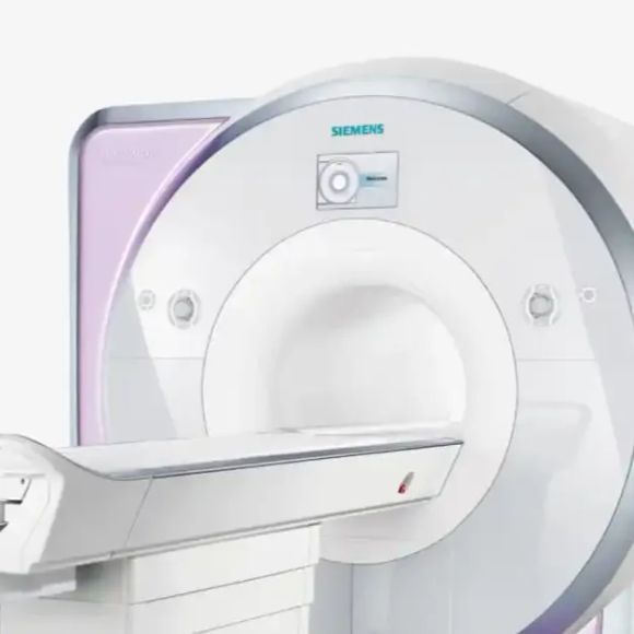
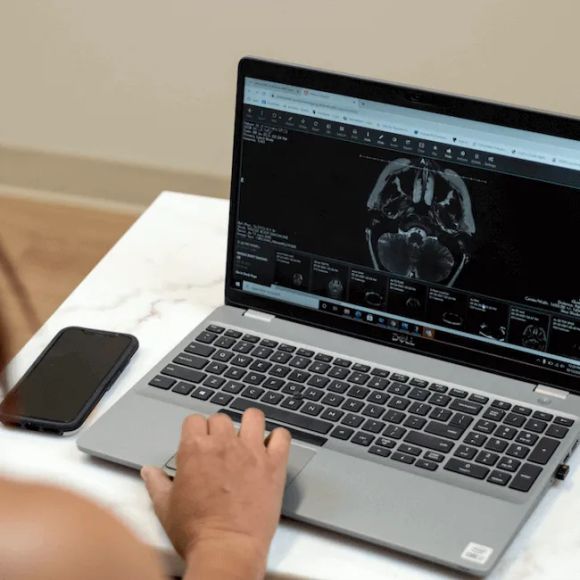
How MRI technology works
Magnetic Resonance Imaging uses powerful magnets and radio waves to create detailed pictures of organs and tissues inside your body. Unlike X-rays or CT scans, MRI does not use ionizing radiation. The machine generates a strong magnetic field that temporarily aligns hydrogen atoms in your body. When radiofrequency pulses disturb this alignment, the atoms emit signals that computers convert into cross-sectional images [1].
This technology excels at showing soft tissues. Doctors can distinguish between normal and abnormal tissue with remarkable clarity. The images reveal details about the brain, spinal cord, joints, muscles, and internal organs that other imaging methods cannot match.
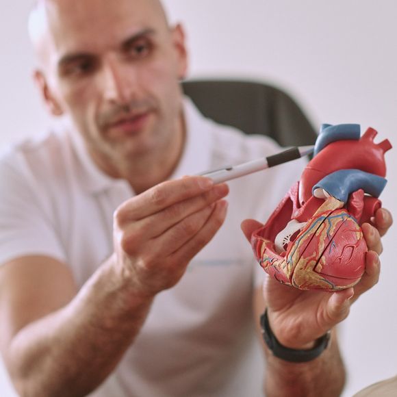
Clinical uses and diagnostic power
Doctors use MRI to diagnose hundreds of conditions. In neurology, it detects brain tumors, strokes, multiple sclerosis, and traumatic injuries. Cardiac MRI evaluates heart structure and function, revealing damage from heart attacks or congenital problems [2]. Orthopedic MRI visualizes torn ligaments, cartilage damage, and spinal disc herniations with precision.
The absence of radiation makes MRI particularly valuable for children and for monitoring conditions over time. You can undergo multiple scans without cumulative radiation exposure. This safety profile supports its use in preventive screening programs for high-risk individuals.
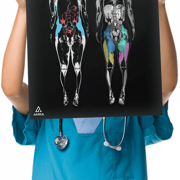
Whole-body MRI screening
Whole-body MRI screening is gaining attention in longevity medicine. These comprehensive scans can detect early cancers, aneurysms, and other serious conditions before symptoms appear. Research suggests whole-body MRI may identify tumors smaller than one centimeter [3].
However, screening carries trade-offs. MRI detects many incidental findings, abnormalities that may never cause problems. These findings can trigger additional testing, biopsies, and anxiety. The benefits of early detection must be weighed against the risks of overdiagnosis and overtreatment.
Safety and limitations
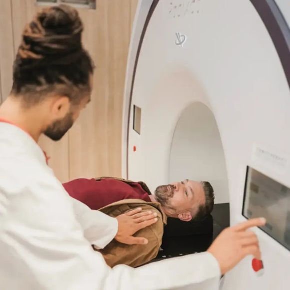
Most people tolerate MRI well. The procedure is painless, though the machine produces loud knocking sounds. Claustrophobia affects some patients, but newer wide-bore machines and mild sedation help. You must remove all metal objects before scanning because the magnetic field can attract them [4].
Certain implants like pacemakers or cochlear devices may prevent MRI. Gadolinium contrast dye helps visualize some structures but requires caution in people with kidney problems. Tell your doctor about any implants, pregnancy, or kidney disease before scheduling.
References
Request ear protection
Remove all metal objects
Disclose all implants and devices
Stay hydrated before contrast scans
Bring prior imaging for comparison
Is MRI safe?
How long does an MRI scan take?
What is gadolinium contrast dye?
Can I have an MRI if I am claustrophobic?
#397 ‒ Endometriosis and adenomyosis: diagnosis, fertility, reproductive aging, and emerging treatments | Renato Tomioka, M.D., Ph.D.
#396 ‒ Breast cancer screening: understanding risk, deciding when to start and how often to screen, and choosing the right imaging strategy
#388 — Prostate cancer screening: why current PSA guidelines are failing men and how modern tools improve early detection and save lives
#386 - Aging clocks—what they measure, how they work, and their clinical and real-world relevance
AMA #78: Longevity interventions, exercise, diagnostic screening, and managing high apoB, hypertension, metabolic health, and more
Lower back pain: causes, treatment, and prevention of lower back injuries and pain | Stuart McGill, Ph.D. (#287 rebroadcast)
No discussions yet
Be the first to start a discussion about MRI.
Related Brands
PreventicsOne
PreventicsOne offers comprehensive preventive, cardiology, and sports medicine services to ensure optimal health.
Aeon
Aeon offers comprehensive health checks with a 60-minute full-body MRI and blood analysis for early detection.

Ahead Health
Ahead Health is a Zurich-headquartered preventive health platform that combines full-body MRI scans, deep blood panels, and AI-driven analysis to detect 500+ potential health risks—empowering longevity seekers to take a proactive path toward lifelong wellness.

Prenuvo
Prenuvo is a healthcare company specializing in advanced whole-body MRI scans designed to detect early-stage cancers and other serious conditions before symptoms appear.
Ezra
Ezra offers full-body MRI screening services designed to detect potential cancer and over 500 conditions across 13 organs in just one hour. Their service is focused on early detection, helping individuals catch cancer early when it is most treatable.
Preventicum
Preventicum is a specialized medical center offering interdisciplinary clinical services in internal medicine, cardiology, gastroenterology, and radiology.
simonONE
simonONE offers a preventative annual body MRI scan service designed to detect potential health issues before they become serious. The service provides a comprehensive health check-up using advanced imaging technology to monitor your body's condition and support early intervention.
Amra Medical
Amra Medical is a leading provider of advanced medical imaging solutions, specializing in body composition analysis through MRI technology.

With Sequel
Sequel is an AI-powered longevity assistant that integrates your health data—including blood labs, MRI and DEXA scans, supplements, and pharmaceuticals—to provide personalized insights. It ensures data privacy by processing information locally on your device, offering both a fully local model and an advanced version power
Hooke
HOOKE London offers personalized health assessments and memberships, utilizing advanced diagnostics and preventive strategies to enhance longevity and overall well-being.